Saturday, October 1, 2016

GI Bleeding in Children


Blood in the vomit.

Blood in the stool.

Blood in the diaper.

How far do I go in my investigation?

What do I really have to worry about?


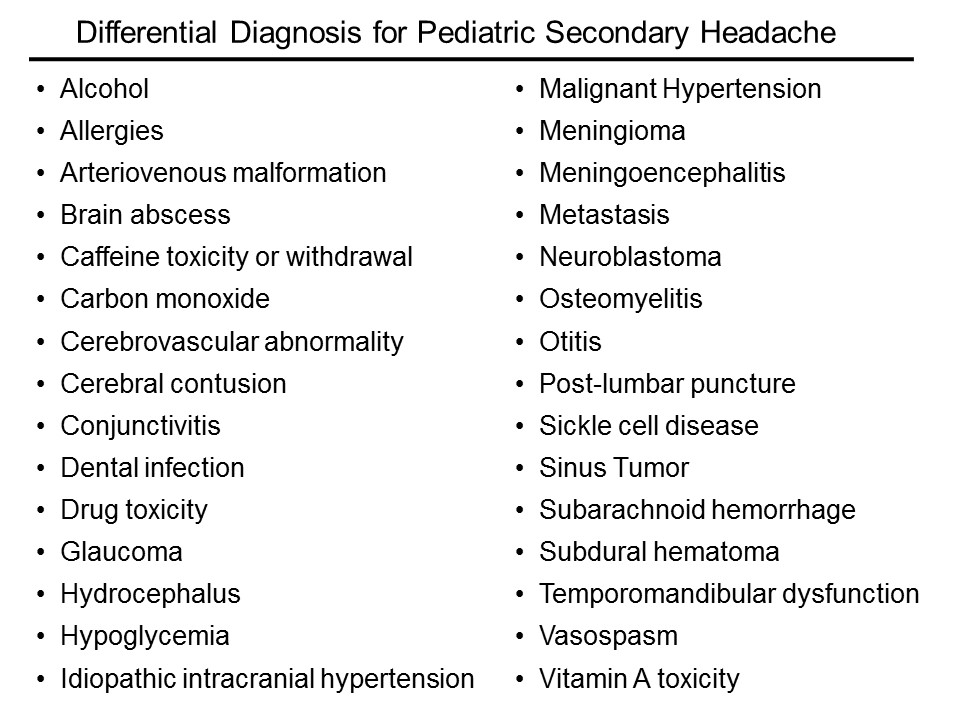
The differential diagnosis of GI bleeding in children is broad.

(Here is the complete differential diagnosis)

In the ED, we can simplify by categorizing by age and appearance.
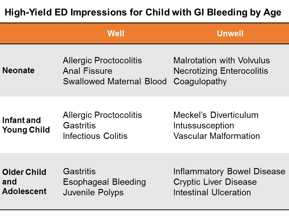
 gi-bleeding-by-age_horeczko


Neonates

GI bleeding in the neonate (less than one month of age) is serious until proven otherwise.

Well appearing?

If this in obvious anal fissure, then no further work-up is necessary.  Counsel on proper feeding and follow-up.

Evaluate for potential swallowed maternal blood by examining mother with a chaperone, then perform the Apt test.

Consider allergic proctocolitis if the child is well.  Counsel the breastfeeding mother on diet modification.  If formula fed, the child should feed through thus until the primary care physician decides whether to start the sticky process of changing up formulas.

If unclear, consider a complete blood count and/or further work-up and admission if unwell.

Ill Appearing?

The three most dangerous diagnoses in the neonate are necrotizing enterocolitis, malrotation with volvulus, and inherited coagulopathy.  It is important to note that 15% of necrotizing enterocolitis occurs in full-term babies; malrotation can present simply in shock, without initial overt bleed.  Inherited conditions may not be known to the family early on, as they have not yet heard back from the neonatal screening done at birth.

Pitfalls in the neonate and infant

Genitourinary bleeding; hematuria; or uric acid crystals: the classic fake out here is the orange or pink stained diaper – that is actually residue from deposits of uric acid crystals in the urine, an almost always benign phenomenon in which the concentrated crystals oxidize and stain the diaper, frightening the parents.

Think -- pink stain, without clot:

Image result for uric acid crystals diaper


Infants and Young Children

Well appearing?

Through the first year to age 5, things like infectious colitis and gastritis are common.

Ill appearing?

Think about intussusception, cryptic liver disease, or esophageal bleeding. Check the skin – is that a dark purple palpable rash on the buttocks? Think Henoch-Schoenlein purpura.

Focus: Meckel's Diverticulum

Meckel’s diverticulum is the most common congenital malformation of the GI tract, and the most common cause of GI bleeding in the toddler.  It is a remnant of the omphalomesenteric tract – it came from a long tube that once connected the yolk sac to the lumen of the midgut.  A stranded island of gastric tissue secretes acid in the intestine, where it doesn’t belong.  Sometimes these islands never cause much trouble.

When it does present itself, a Meckel’s diverticulum usually follows the rule of twos:

Presents by age 2
Affects 2% of the population
Often 2 inches in length
May include 2 types of mucosa
Found within 2 feet of the ileocecal valve.

Not actively bleeding: technetium-99 pertechnate scintigram (Meckel’s scan).

Actively bleeding: radio-labeled red blood-cell scan (resuscitate and call your surgeons!)

Pitfalls in the infant and young child

Epistaxis; food-related misadventures


Older Child and Adolescent

Well appearing?

Mallory-Weiss tears after forceful vomiting; trivial hemoptysis after viral symptoms; pill esophagitis in the child is just learning to swallow medications.  Always consider foreign body ingestion.

Ill Appearing?

Varices from cryptic liver disease; hemorrhagic gastritis; vascular malformation, such as a Dieulafoy lesion, where a tortuous small artery ends just superficial to the gastric mucosa, and can erode through and erupt.

Focus: Inflammatory Bowel Disease

Approximately a quarter of patients with inflammatory bowel disease (IBD) -- both Ulcerative Colitis and Crohn disease – will present by age 20.  Children and adolescents may present with the classic symptoms of IBD: abdominal pain, weight loss, bloody diarrhea, but many present atypically with isolated signs like poor growth, anemia, or delayed puberty.

You may also suspect IBD in the child with other extra-intestinal symptoms like oral ulcers, clubbing, erythema nodosum, jaundice, or hepatomegaly.

On history and physical examination, you may get one of three cardinal presentations

Fatigue, history of anemia, in a stable child who comes to the ED with bloody diarrhea

Chronic diarrhea, chronic abdominal pain, and poor weight gain or weight loss

A fulminant presentation, with severe abdominal pain, frankly bloody stools, tenesmus, fever, leukocytosis, and hypoalbuminemia.

On exam, look for general appearance, glossitis from B2 deficiency, hair loss and brittle nails form protein loss, purpura (from vitamin C and vitamin K deficiencies).  Look for evidence of episcleritis or uveitis.  Listen for rubs as in pericarditis.  Do a good abdominal exam, especially looking for hepatomegaly. Perirectal skin tags are not uncommon. Children with IBD may form urinary calculi form oxalate crystal deposition. Do a thorough skin and neurologic exam.

Treatment for both ulcerative colitis and Crohn’s disease is similar.

Induction therapy: children with mild disease get aminosalicylates; those with moderate disease get steroids; and those with severe disease get cyclosporine.

Maintenance regimens to prevent relapse include aminosalicylates, mercaptopurine, and azathioprine.

Surgical treatments for refractory colitis include an ileal pouch and anal anastomosis – also called a J pouch, a type of neorectum created surgically by folding loops of ileum back on themselves and stitching them together to create a larger rectal reservoir where the rectum once was.  A neorectum allows the child to have voluntary control of his stools again.

Stabilizing the Pediatric GI Bleed

Life-threatening GI bleeding in children is, thankfully, rare, but we have to be prepared.

Give blood for compensated shock, prepare for massive transfusion if giving more than 40 mL/kg total blood products.

Differing Etiologies: adults and children

The reasons for upper GI bleed in adults are vastly different from those of children.  In adults, mostly the life-threats are due to liver disease, varices, or hemorrhagic gastritis.

In children, critical upper GI bleed is often secondary to critical illness (hemorrhagic gastritis or stress ulcer), or vascular malformation.  Critical lower gastrointestinal bleeding may be from Meckel's diverticulum or other congenital angiodysplasia.

Endoscopy

Get your patient urgent endoscopy as soon as possible after arrival if there is active bleeding. Otherwise, according to the Belgian guidelines, stable children may have endoscopy within the first 24 hours of hospitalization. The reported efficacy of endoscopy for controlling upper GI bleeding in children is approximately 90%.

Miscellaneous

Nasogastric tube? No routine role (unreliable to rule out or stratify upper GI bleed).

Proton pump inhibitor? No good data, but no major common contraindications.

Octreotide, vasopressin, broad-spectrum antibiotics?  May use adult data to extrapolate in the proper etiologic context.

General Advice for the Brisk GI Bleed in Children

This is a rare, but potentially life threatening situation, so anticipate how the child can decline, and get your team assembled: your pediatric intensivist, gastroenterologist, and surgeon – especially if we can’t ge the upper GI bleed to abate with endoscopy. The sooner you activate the team, the better.

Summary

The broad differential diagnosis may be paralyzing, and frustrating, since much of it we cannot discern in the ED.

Consider actionable, high-yield etiologies based on age and appearance.

Neonates

Well appearing?  Think rectal fissure, maternal blood, or allergic proctocolitis
Ill appearing?  Think necrotizing enterocolitis, malrotation, or an inherited coagulaopathy.

Infants, and Young Children

Well appearing? Think infectious colitis and gastritis.
Ill appearing? Think intussusception and Meckel’s diverticulum.

Older Children and Adolescents

Well appearing?  Think Mallory Weiss tear from vomiting, or gastritis
Ill appearing?  Think metabolic, cryptic liver disease, or inflammatory bowel disease.

For everyone – a careful history and a good physical exam will point you tto the etiology, or risk-stratify for further outpatient evaluation and management.

References

Bozic MA, Puri K, Molleston JP. Screening and Prophylaxis for Varices in Children with Liver Disease. Curr Gastroenterol Rep. 2015 Jul;17(7):27.

Chaïbou M, Tucci M, Dugas MA, Farrell CA, Proulx F, Lacroix J. Clinically significant upper gastrointestinal bleeding acquired in a pediatric intensive care unit: a prospective study. Pediatrics. 1998 Oct;102(4 Pt 1):933-8.

Colle I, Wilmer A, Le Moine O, Debruyne R, Delwaide J, Dhondt E, Macken E, Penaloza A, Piessevaux H, Stéphenne X, Van Biervliet S, Laterre PF. Upper gastrointestinal tract bleeding management: Belgian guidelines for adults and children. Acta Gastroenterol Belg. 2011 Mar;74(1):45-66.

Garcia-Tsao G, Bosch J. Varices and Variceal Hemorrhage in Cirrhosis: A New View of an Old Problem. Clin Gastroenterol Hepatol. 2015 Nov;13(12):2109-17.

Kaya A, Toyran M, Civelek E, Misirlioglu E, Kirsaclioglu C, Kocabas CN. Characteristics and Prognosis of Allergic Proctocolitis in Infants. J Pediatr Gastroenterol Nutr. 2015 Jul;61(1):69-73.

Lacroix J, Nadeau D, Laberge S, Gauthier M, Lapierre G, Farrell CA. Frequency of upper gastrointestinal bleeding in a pediatric intensive care unit. Crit Care Med. 1992 Jan;20(1):35-42.

Osman D, Djibré M, Da Silva D, Goulenok C. Management by the intensivist of gastrointestinal bleeding in adults and children. Ann Intensive Care. 2012 Nov 9;2(1):46.

Owensby S et al. Diagnosis and Management of Upper Gastrointestinal Bleeding in Children. J Am Board Fam Med. 2015; 28(1):134-145.

Rosen MJ et al. Inflammatory Bowel Disease in Children and Adolescents. JAMA Pediatr. 2015 Nov;169(11):1053-60.

Rufo PA, Bousvaros A. Current therapy of inflammatory bowel disease in children. Paediatr Drugs. 2006;8(5):279-302.

Srygley FD, Gerardo CJ, Tran T, Fisher DA. Does this patient have a severe upper gastrointestinal bleed? JAMA. 2012 Mar 14;307(10):1072-9.

Thomson MA, Leton N, Belsha D. Acute upper gastrointestinal bleeding in childhood: development of the Sheffield scoring system to predict need for endoscopic therapy. J Pediatr Gastroenterol Nutr. 2015 May;60(5):632-6.

This post and podcast are dedicated to Carlo D'Apuzzo, MD  for his creativity, innovation, and dedication to the highest standards of emergency care.  Le tue pillole sono buona medicina. Grazie, Carlo per tutto quello che fai per il mondo #FOAMed.


150px-WikEM_app_Logo


GI Bleeding in Children

Powered by #FOAMed -- Tim Horeczko, MD, MSCR, FACEP, FAAP


Check out this episode!

Thursday, September 1, 2016

Pediatric Headache: Some Relief for All


Seemingly vague, but potentially dangerous...

common, but possibly with consequences...

...or maybe just plain frustrating.

Let's talk risk stratification, diagnosis, and management.


Primary or Secondary?

We can make headache as easy or as complicated as we like, but let's break it down to what we need to know now, and what the parents need to know when they go home.

Primary headaches: headaches with no sinister secondary cause – like tension or migraine – are of course diagnoses of exclusion (cluster headache is exceedingly rare in children).

Secondary headaches: headaches due to some underlying cause -- are what we need to focus on first.

The list of etiologies is vast; here is just a sampling:
Horeczko_Podcast_Differential Diagnosis Headache

How do I sort this out?

Ask yourself three main questions:

Is it a tumor?

Is it an infection?

Is it a bleed?

Is it a tumor?

Some historical features are high-yield in screening for signs or symptoms consistent with a space occupying lesion.

Progression and worsening of symptoms over time

Associated vomiting

Pain only in the occiput

Headache that is worse with Valsalva – ask if coughing, urinating, or defecating affects the headache

Does this headache wake the child from sleep?

Is it worse in the morning just after getting up?

Conversely, the absence of some historical features may increase suspicion of a space-occupying lesion

No family history of migraine

No associated aura with the headache.

Who needs neuroimaging?

The short answer is, if the child has an abnormal exam finding, then obtain a non-contrast head CT in the ED.  If you’re worried enough to get imaging, then you should not feel great about sending him to an expedition to MRI.

The reassuring point is that for a child with a normal neuro exam, we have time to figure this out. For the recurrent headache, outpatient MRI really is the way to go if at all possible – not only do we forgo unnecessary radiation, but MRI is more likely to reveal the cause – or rule out the concern.

Medina et al. in Pediatrics reported on children with headache suspected of having a brain tumor. They stratified patients into low, intermediate, and high risk, based on clinical predictors from the history and physical. All had imaging. They then calculated probability of tumor in each group.

The low risk group had a 0.01% probability of tumor. The intermediate group 0.4%, and the high-risk group had only a 4% probability of tumor. The take-home message is that in the stable patient with a normal neurologic exam and no red flags, time is on our side.

The American Academy of Neurology's most recent guidelines, published first in 1994 and revised in 2004.

1. Neuroimaging on a routine basis is not indicated with recurrent headaches and a normal neurologic exam

2. Neuroimaging should be considered in children with an abnormal exam.

3. Neuroimaging should be considered in children with recent onset of severe headache, change in the type of headache, or associated features that suggest neurologic dysfunction

Is it an infection?

This is nothing new: if you think you need to perform a lumbar puncture, then you’re right. Go after the diagnosis when it meets your threshold for testing.

The difficulty is in the child who just has a headache, plus or minus symptoms that may be viral syndrome.

Dr Curtis et al. in Pediatrics did a systematic review of Clinical Features Suggestive of Meningitis in ChildrenIn the history, only obvious features were helpful in this study: bulging fontanel in the infant or neck stiffness in the older child.  Both increased the likelihood of meningitis by 8-fold.

In the physical examination, the only reliable predictors in this study were poor general appearance or a change in behavior.

You will catch those cases, because you would have tuned into meningitis early on -- especially in the unvaccinated.

What about all-comers with fever and headache? The presence of a high fever (so greater than 40 °C) only conferred a positive likelihood ratio of 2.9, only marginally predictive. Reassuring is that for temperatures less than 40 °C, the LR was 1 for meningitis.

In other words, a fever less than 40 °C was just as likely to be present with or without meningitis.

Is it a bleed?

Does this child have some underlying disorder? For example, sickle cell disease, hypertension, rheumatologic disease, or some other endocrine or metabolic disease, such as a mitochondrial disorder?

In chronically ill children, consider cerebral sinus venous thrombosis, vasculitis, ischemia, or hemorrhage.

Arteriovenous malformation (AVM) is the hemorrhage we fear the most.

We really don’t know enough about arteriovenous malformations in the brain to say what is the typical presentation. They may be completely asymptomatic, until they rupture. Even the headache presentation is variable.

Think, headache PLUS.

New headache plus…vomiting.

Headache plus…it’s unilateral and new for the patient.

Headache plus…a new seizure.

Headache plus…focal neuro deficits, that may be transient, due to a vascular steal phenomenon.

Two illustrative cases of arteriovenous malformation:

1. An eleven-year-old girl presents to the ED with new headache, nausea, and vomting in the morning, then had a generalized seizure later that day, and presents with a low GCS. She was intubated, CT confirmed the AVM. She had a right frontal intraparenchymal bleed with midline shift. She underwent clot evacuation and extirpation of the intertwined arteries and veins.

2. A nine-year old girl presented to the ED with headache for two days, constant, then one day of nausea and vomiting. On presentation, she was altered, and had slow-reacting pupils. She also underwent evacuation, and only on histopathology did they find a single, arterialized vein.

Primary Headache: Presumptive Impression

Tension headaches are the most common in children and adults. As in adults, the tension headache is band-like, pressure, tighetening, and often associated with muscle aches in the neck and shoulders.

Find out how often they occur, and whether there is any pattern of worsening symptoms, or if the symptoms seem to be related to sleep hygiene, video games, too much digital screen time. Also, screen for lack of exercise, poor diet, stress, and all of the other good questions you usually ask.

Treat the cause or counsel about lifestyle modification, and offer PO hydration and an NSAID, like ibuprofen or acetaminophen (paracetamol).

Non-pharmacologic techniques like heat packs, rest, stress relief, and a little TLC always help. Be careful not to encourage overreacting to the headache – sometimes we see a pattern of headache, attention, and more headache that can take root. Also look for overuse of medications, which may be the culprit in up to 50% of chronic headaches. Taking NSAIDs 3 or more times per week is associated with medication-induced headache, or cephalalgia medicamentosa.

We often fail to identify migraine headaches in children in the ED, likely for two reasons: prevalence of migraine increases with age, and children don’t present exactly like adults.

Stewart et al. in Neurology, report a prevalence of migraine in children that increases with age: 3 to 7 years of age was 2%; 7 to 11 years of age, 7%; and 11 to 20 years of age, 20%

Pearl: migraines are most commonly bilateral and temporal in children.  They resemble "adult" tension headaches, but are much more severe.

We may not be able to sort this out in the ED.  The point here is that migraines in children are more common that we may expect, and they can interfere with school performance, with social development, or even with family dynamics and overall stress burden.

Primary Headache Diagnosis: Not (Usually) "Our Thing"

You noticed that we treated before we knew exactly the etiology; such is Emergency Medicine.

We may not be able to make a specific, definitive primary headache diagnosis in the ED, but we should be aware of the criteria to help counsel patients and families.

Tension headache is the most common, but it requires multiple, similar episodes:Horeczko Podcast Tension Headache

Migraine headache (without aura) requires less episodes, but more specific features:

Slide3

An aura is a fast-pass to diagnosis of migraine:Horeczko Podcast Migraine with Aura


Primary Headache Management

So how do we treat primary headaches? If you feel this is a mild tension headache, fluids by mouth and a simple NSAID are probably all that is needed, in addition to a heaping dose of reassurance.  Ibuprofen (10 mg/kg/dose q 6h, up to 600 mg) for a short course has the most evidence basis.  Acetaminophen (paracetamol) (15 mg/kg/dose q6 h) for a short course may also be given.

Abortive treatments with the triptans may have been tried at home, but if they are coming to see us, we are past the point where triptans will be helpful.

For the primary headache that is resistant to NSAIDs, IV therapy may be considered.

If you’re going for IV, a nice evidence-based migraine cocktail is the following:

1. A bolus of 20 ml/kg of normal saline, up to a liter

2. Ketorolac (0.5 mg/kg; max, 30 mg)

3. Diphenhydramine (2 mg/kg; max, 50 mg)

4. Prochlorperazine (0.1 mg/kg; max, 10 mg)

Dr Kaar et al. in Pediatric Emergency Care evaluated the safety and efficacy of their institution’s standardized pediatric migraine practice guideline in the emergency department, which used ths cocktail, based on the best evidence available. In their retrospective chart review, they found the average visual pain scale drop from 7.8 to 2.1

There were no adverse events reported.

So, really you can treat children with migraines very similarly to adults.

Other treatments on the horizon (still under investigation) in children include IV adjuncts such as magnesium, valproic acid, and dexamethasone.

Aftercare and Recurrence Prevention

For everyone who is going home, take just a moment to talk about the importance of sleeping well, eating well, getting exercise, limiting digital screen time, and trying to improve ways of dealing with stress.

When all else fails, and the parent has “heard it all”: get them started on a headache diary.

Take a piece of paper, fold it in half, and start a template for them to work on in a spiral notebook.  Start a sample entry for them, with the date and time the headache started, what it felt like, what was happening just before, what made the headache better, any dose of medications given, how long it lasted, and what the patient did after. There are even free apps that will track the headache pattern.

This is the first thing a neurologist will start them on – and it’s sometimes a selling point to the parent that the time spent waiting for a referral to a neurologist is not waste – they will actually be in better shape and can move things along faster.  It also gives them some sens of control of what can be a draining situation.

Summary and Mental Road Map

If you were thinking meningitis or acute bleed, especially with fever or meningismus, get a CT first if you see signs of increased intracranial pressure, or if there is an abnormal neuro exam. Otherwise go straight to the lumbar puncture (LP).

In the afebrile child with a normal exam, give symptomatic relief, briefly counsel them, and arrange for follow-up.

In the afebrile child with an abnormal exam, obtain a CT in the ED. If negative, either admit for MRI if you are still concerned, or consider LP for idiopathic intracranial hypertension (pseudotumor cerebri).

Talk with parents early about expectations, and offer them some friendly advice on prevention. Refer patients to the primary care provider or neurologist if the presentation is more involved.

After a good history and physical examination in the ED that results in no red flags, we have time on our side. Help the family through the process by explaining the next steps and what can be done in the meantime. Compassion and a plan: sometimes these are our most powerful allies.


References

Ajiboye N et al. Cerebral Arteriovenous Malformations: Evaluation and Management. Scientific World J. 2014; vol 2014.

Bachur RG, Monuteaux MC, Neuman MI. A comparison of acute treatment regimens for migraine in the emergency department. Pediatrics. 2015 Feb;135(2):232-8. doi: 10.1542/peds.2014-2432.

Chiappedi M, Balottin U. Medication overuse headache in children and adolescents. Curr Pain Headache Rep. 2014 Apr;18(4):404. doi: 10.1007/s11916-014-0404-9.

Curtis S, Stobart K, Vandermeer B, Simel DL, Klassen T. Clinical features suggestive of meningitis in children: a systematic review of prospective data. Pediatrics. 2010;126(5):952-60.

Gonzalez LF, Bristol RE, Porter RW, Spetzler RF. De novo presentation of an arteriovenous malformation. Case report and review of the literature. J Neurosurg. 2005 Apr;102(4):726-9.

Kaar CR, Gerard JM, Nakanishi AK. The Use of a Pediatric Migraine Practice Guideline in an Emergency Department Setting. Pediatr Emerg Care. 2016 Jul;32(7):435-9. doi: 10.1097/PEC.0000000000000525.

Lewis DW, Ashwal S, Dahl G, Dorbad D, Hirtz D, Prensky A, Jarjour I; Quality Standards Subcommittee of the American Academy of Neurology; Practice Committee of the Child Neurology Society. Practice parameter: evaluation of children and adolescents with recurrent headaches: report of the Quality Standards Subcommittee of the American Academy of Neurology and the Practice Committee of the Child Neurology Society. Neurology. 2002 Aug 27;59(4):490-8.

Lewis D, Ashwal S, Hershey A, Hirtz D, Yonker M, Silberstein S; American Academy of Neurology Quality Standards Subcommittee; Practice Committee of the Child Neurology Society.Practice parameter: pharmacological treatment of migraine headache in children and adolescents: report of the American Academy of Neurology Quality Standards Subcommittee and the Practice Committee of the Child Neurology Society.Neurology. 2004 Dec 28;63(12):2215-24.

Richer L, Billinghurst L, Linsdell MA, Russell K, Vandermeer B, Crumley ET, Durec T, Klassen TP, Hartling L. Drugs for the acute treatment of migraine in children and adolescents. Cochrane Database Syst Rev. 2016 Apr 19;4:CD005220.

Stewart WF, Lipton RB, Celentano DD, Reed ML. Prevalence of migraine headache in the United States. JAMA. 1992;267:64-69.

Tascu A et al. Spontaneous intracranial hemorrhage in children – ruptured lobar arteriovenous malformations: report of two cases. Romanian Neurosurgery. 2015; 29(23) 1: 85-89.

This post and podcast are dedicated to Mark Wilson, PhD, BSc, MBBChir, FRCS(SN), MRCA, FIMC, FRGS for his #FOAMed generosity, candor, humility, and dedication to the care of the acutely ill and injured. Thank you.


150px-WikEM_app_Logo

Pediatric Headache

Powered by #FOAMed -- Tim Horeczko, MD, MSCR, FACEP, FAAP


Check out this episode!

Monday, August 1, 2016

Subcutaneous Rehydration


Have you ever been in any of these situations?

⇒   You have a stable child who just needs fluids, but no laboratory tests
⇒   You’ve tried PO hydration, to no avail, despite anti-emetics
⇒   You’re poking the stable, but dehydrated child repeatedly without success

What now?

Hypodermoclysis, otherwise known as subcutaneous rehydration.

[Insert Player]
Clysis comes from the same Greek word that “a flood” – hypodermoclysis refers to flooding the subcutaneous space with fluid, so that it can be absorbed systemically.

Sound far-fetched?

Well, it turns out, what is old is new again.

In 1913, Dr Day first described this technique for a child with severe diarrhea who could not tolerate fluids by mouth. Hypodermoclysis then began to gain popularity with a peak of use in the 1940s, until an innovative breakthrough in 1950. Dr David Massa, a resident anesthesiologist at the Mayo clinic, invented the first catheter-over-needle apparatus.

With increasing safety and ready access of IV catheters, IV quickly overshadowed SC.

The subcutaneous route of hydration has also been used effectively in geriatric and palliative care for decades, and it is only now beginning to gain popularity again in its original population: children.

So, how does it work?

In a nutshell, you place a butterfly needle or angiocatheter in the subcutaneous space and you run fluids into it. The tissues quickly absorb the fluids, making them available systemically. That’s it. Everything else is just finesse.

The ideal candidate for hypodermoclysis is the stable patient, with mild to moderate dehydration who fails a trial of fluids by mouth, or who needs a bridge to gaining IV access later, after a slow subcutaneous fluid bolus is given.

Ok, so how do you do it?

Place a topical anesthetic cream, such as EMLA, cover with occlusive dressing (IV dressing), wait 15-20 min

"Pinch an inch" of skin anywhere, but the most practical site in young children is between the scapulae

Insert a 25-gauge butterfly needle or 24-gauge angiocatheter (preferred by the author), secure

Inject 150 U hyaluronidase SC, if available

Infuse 20 mL/kg isotonic solution over one hour, repeat as needed or use "bolus" as bridge to IV access

You can set the line to gravity, and if it is dripping in, you may leave it be. If you see a very slow drip by gravity, or worse, nothing is dripping, you can set the line on a pump, to deliver up to 20 mL/kg over an hour. Infusion at this rate optimizes the balance we want in minimal discomfort while maximizing the flow rate.

This is not a “bolus” in the true sense – but then, when you compare it to the alternative – like IV therapy – and we see a time and cost savings.  Dr Mace and colleagues in the American Journal of Emergency Medicine report substantially decreased cost and ED length of stay when comparing the material and human resources needed to place an IV in a squirmy young child, compared with a simple subcutaneous stick.

There will be swelling

There will be swelling – that is the goal. It is really painless, and your patient may lie down on his back with the pump going – it is actually pretty comfortable for most children and adults to do.

Hypodermoclysis_Photo_HoreczkoHere’s a tip – since there will be swelling, we want to be careful about how we secure the line, so how you tape it down to the skin is important – we want to avoid a pulling sensation, which can be the beginning of the end of the tolerance for the procedure.  Cover that with an occlusive dressing, as you would an IV site. The footprint of the occlusive dressing is relatively small, so it will travel up on top of the subcutaneous mound you’re creating. As the line exits the occlusive patch, place a thin layer of gauze between the skin and the IV tubing, so that the tubing doesn’t press into the skin. Then—as far away from the puncture site as possible—tape it down securely. The idea is not to tape on the growing mound itself, because the mound may pull at the anchored skin and set a nuclear chain reaction of annoyance and restlessness – and potentially a failed procedure.

The swelling will look indurated, a pinkish red.  It’s not an allergic reaction: even with the old preparations of hyaluronidase, allergic reactions were rare, and now they are very rare with the recombinant preparation. It is supposed to swell and look ugly. The subcutaneous tissues will swell to a point where you have a steady state fluid administration rate, and as soon as you stop the infusion, the remaining fluid will start to subside as it is absorbed.

A Bridge to IV Therapy?

Kuensting et al. in the Journal of Emergency Nursing in 2013 compared subcutaneous fluid infusion with intravenous fluid infusion in children with difficult IV access. They found the mean time from order entry to subcutaneous fluid infusion to be 20 min, compared to the failed IV access group with an average infusion start time of 1.5 hours. The latter group eventually received subcutaneous fluids.  The investigators also found a shorter ED length of stay in the subcutaneous group.

In the same study, a subgroup received subcutaneous fluids initially, and later an IV. They found a trend in ease of IV access after subcutaneous fluid therapy. In other words, if your little patient with difficult IV access is hemodynamically stable and amenable to a bolus over an hour, you may choose to start with hypodermoclysis and reevaluate.

Predicting Difficult IV Access in Children

Much has been studied and written about the predictors of difficult IV access in children. The most often cited are: age < 3 years, weight less than 5 kg, prematurity, obesity, and darker skin tones, where the contrast of vein to skin may not be so apparent.

The three main predictors of the score validated by Riker et al. in Annals of Emergency Medicine include the most practical and universal of features: vein palpability, vein visibility, and patient age.

If you’re anticipating difficult IV access in the child who can stand to wait an hour for a slow bolus, you may start with the subcutaneous route to get those veins plumper and more visible, to improve your chances of IV access in the very near future.

Medications via Subcutaneous Route

Certain medications have been used safely via subcutaneous infusion; always check dose, rate, and compatibility.

SC Medications_Horeczko

Compatibility of Subcutaneous Medications


What about catheter size?

You don’t need to use larger needles or angiocathters for older children, adolescents or adults. A 25-gauge butterfly or 24-gauge angiocatheter works well from an infant to an elder. In one study of adults, a half a liter of saline was infused by gravity via a 24-gauge catheter. With IVs, the shorter and larger the bore, the faster the infusion.

In subcutaneous infusion, it is not the size of the catheter, but the osmotic gradient that determines the rate of absorption.

What if I don't have that fancy hyaluronidase?

It’s actually increasingly readily found – and available in generic form. If you have it, please use it – it will make a believer out of you and others.

Hypodermoclysis will work without hyaluronidase – the process of subcutaneous rehydration just takes a lot longer to work. In a double-blind cross-over trial Thomas et al. in 2007 compared subcutaneous administration of lactated ringer’s solution by gravity with and without hyalurondase. The hyaluronidase group received their fluids 5 times faster. The average rate of the hyaluronidase group was 382 mL/h versus the fluid only group, who did not receive hyalurinodase; they were substantially slower, at 82 mL/h. It’s worth using if you have it, but still potentially useful if you don’t.

Recap: Supplies

√    EMLA or any topical anesthetic used for intact skin, placed as soon as the decision is made
√    A 25-gauge butterfly needle or 24-gauge angiocatheter
√    IV tubing, gauze to pad, tape to anchor
√    150 U hyaluronidase, the same dose, regardless of age or size
√    Isotonic fluids – you can start with 20 ml/kg
√    And finally a well informed team made up by the patient, the parents, and your staff, so that everyone knows what to expect for a successful subcutaneous fluid administration.

References

Allen CH, Etzwiler LS, Miller MK, Maher G, Mace S, Hostetler MA, Smith SR, Reinhardt N, Hahn B, Harb G; INcreased Flow Utilizing Subcutaneously-Enabled Pediatric Rehydration Study Collaborative Research Group. Recombinant human hyaluronidase-enabled subcutaneous pediatric rehydration. Pediatrics. 2009 Nov;124(5):e858-67.

Bruno VG. Hypodermoclysis: a literature review to assist in clinical practice. Einstein (Sao Paulo). 2015 Jan-Mar;13(1):122-8.

Cabañero-Martínez MJ, Velasco-Álvarez ML, Ramos-Pichardo JD, Ruiz Miralles ML, Priego Valladares M4, Cabrero-García J. Perceptions of health professionals on subcutaneous hydration in palliative care: A qualitative study. Palliat Med. 2016 Jun;30(6):549-57.

Kuensting LL. Comparing subcutaneous fluid infusion with intravenous fluid infusion in children. J Emerg Nurs. 2013 Jan;39(1):86-91.

Mace SE, Harb G, Friend K, Turpin R, Armstrong EP, Lebel F. Cost-effectiveness of recombinant human hyaluronidase-facilitated subcutaneous versus intravenous rehydration in children with mild to moderate dehydration. Am J Emerg Med. 2013 Jun;31(6):928-34.

O'Hanlon S, Sheahan P, McEneaney R. Severe hemorrhage from a hypodermoclysis site. Am J Hosp Palliat Care. 2009 Apr-May;26(2):135-6.

Remington R, Hultman T. Hypodermoclysis to treat dehydration: a review of the evidence. J Am Geriatr Soc. 2007 Dec;55(12):2051-5.

Riker MW, Kennedy C, Winfrey BS, Yen K, Dowd MD. Validation and refinement of the difficult intravenous access score: a clinical prediction rule for identifying children with difficult intravenous access. Acad Emerg Med. 2011 Nov;18(11):1129-34.

Rouhani S, Meloney L, Ahn R, Nelson BD, Burke TF. Alternative rehydration methods: a systematic review and lessons for resource-limited care. Pediatrics. 2011 Mar;127(3):e748-57.

Smith LS. Hypodermoclysis with older adults. Nursing. 2014 Dec;44(12):66.

Spandorfer PR. Subcutaneous rehydration: updating a traditional technique. Pediatr Emerg Care. 2011;27(3):230-6.

Thomas JR, Yocum RC, Haller MF, von Gunten CF. Assessing the role of human recombinant hyaluronidase in gravity-driven subcutaneous hydration: the INFUSE-LR study. J Palliat Med. 2007 Dec;10(6):1312-20.

Vacha ME et al. The Role of Subcutaneous Ketorolac for Pain Management. Hosp Pharm. 2015 Feb; 50(2): 108–112.

Zaloga GP, Pontes-Arruda A, Dardaine-Giraud V, Constans T; Clinimix Subcutaneous Study Group. Safety and Efficacy of Subcutaneous Parenteral Nutrition in Older Patients: A Prospective Randomized Multicenter Clinical Trial. J Parenter Enteral Nutr. 2016 Feb 17. pii: 0148607116629790. [Epub ahead of print]


This post and podcast are dedicated to Christina L. Shenvi, MD, PhD, for her dedication to excellence in patient care and enthusiasm in #FOAMed, Emergency Medicine, and Geriatric Emergency Medicine.  There are many shared lessons learned in the care of children, elders, and families.  Thank you.

Catch Dr Shenvi on the innovative GEMcast.


150px-WikEM_app_Logo

Subcutaneous Infusion

Powered by #FOAMed -- Tim Horeczko, MD, MSCR, FACEP, FAAP


Check out this episode!

Friday, July 1, 2016

Please STOP LIMPING!


"She won't walk", or "He just looks like he's limping".

So many things can be going on -- how do we tackle this chief complaint?

You’re dreading a big work-up.  You almost want to tell the kid – please, STOP LIMPING...

STOP LIMPING!

S – Septic Arthritis 

The most urgent part of our differential diagnosis. The hip is the most common joint affected, followed by the knee.  Lab work can be helpful, as well as US of the hip to look for an effusion,  but sometimes, regardless of the results, the joint just has to be tapped to know for sure.

T – Toddler’s fracture

This is usually a torque injury when the wobbling toddler pivots quickly or trips and falls.  Toddler’s fractures happen in children 1 to 3 years of age, and occur in the distal 1/3 of the tibia.  Sometimes a cast is needed, but currently there is a new trend in foregoing casting in mild cases.

O – Osteomyelitis

Bacteremia – from any source – can seed into any bone.  It’s not very common, but it happens: approximately 2% of children who present to an ED with limp will have osteomyelitis.  Plain films, ESR, and CRP are a fair screen to start.  For more than the casual concern, MRI is the best modality to evaluate, followed by radionuclide scintigraphy.  Although not the first choice modality, CT can show periosteal changes, such as inflammatory new bone formation or periosteal purulence.

P – Perthes disease

This is the famous Legg-Calvé-Perthes idiopathic avascular necrosis of the hip, usually affecting children from 3 to 12 years. They present with a slow onset pain and with an antalgic gait.  Patients will have trouble with internal rotation and abduction of the hip.  Radiographs may be initially normal.  MRI can show the culprit: decreased perfusion to the femoral head and subsequent necrosis.

L – Limb-Length Discrepancy

Parents may notice that he seems “wobblier” than he should be.  It may be that we are just now appreciating a congenital anomaly.  Get out the paper tape, and measure from the anterior superior iliac spine to the medial malleolus and compare both sides.   Children with limb-length discrepancy only need a non-urgent referral to pediatric orthopedics to look for congenital dysplasia of the hip, or other growth abnormalities.  Some are treated with orthotics.  Surgical options vary.  Epiphysiodesis destroys the growth plate on the unaffected side, which evens out the growth.  Other options are limb-lengthening or limb-shortening procedures.

I – Inflammatory

Transient Synovitis.  This is what we want them to have right?  The typical age is between 3 and 6 years, sometimes just after a URI.  To be comfortable with this diagnosis, we should have considered all of the dangerous diagnoses, the child should be well, afebrile, in minimal discomfort, and he should respond almost completely to an NSAID.  He’s the one running up and down the department after treatment – or just from sheer boredom after observation.

M – Malignancy

Primary bone tumors such as Ewing’s sarcoma or osteogenic sarcoma typically affect older children.  Limping, however, may be a presenting symptom of leukemia.  If you have any suspicion of the general wellness of the child, get a screening CBC, and perhaps a peripheral blood smear.  Whatever you do, make sure you get close follow up for these kids that are on your malignancy radar -- the blast crisis may not have occurred yet – but it can happen hours to days later.

Plain films are insensitive for leukemic involvement of bone but they may show diffuse osteopenia, or metaphyseal bands – symmetrical high-uptake markings around the joint.  They look like stacks of paper within normal bone – you can see them also in anemia, lead poisoning, and other causes.  Also look for periosteal new bone formation, sclerosis, or lysis.

P – Pyomyositis

This usually presents with vague irritability, pain, and fever, and sometimes with a subacute minor trauma.  These children don’t look to well.

Also think about just run-of-the-mill myositis, usually from a viral cause, such as influenza.  Typically the calves are affected and are always tender.  Hydration and supportive therapy are indicated for viral causes.

For bacterial focal pyomyositis, give empiric antibiotics, admit them for major inpatient workup, and think about early surgical consultation if you think you need sepsis source control.

I – Iliopsoas Abscess

Children most often will develop a primary abscess from bacteremia from an unresolved infection.   Adults more commonly form secondary abscesses from Crohn’s disease, post-op complications, a vertebral infection, or even a bad chronic urinary tract infection.  Lest you think this is a dramatic presentation, think again: iliopsoas abscesses present also with vague symptoms of back, flank, abdominal, or hip pain, sometimes with fever.  The median time from symptoms to diagnosis in children is a whopping 20+ days, according to one study.  If iliopsoas abscess is starting to get your attention, get the CT or MRI.

N – Neurologic

Not to scare you, but children do have strokes; unlike adults, half are hemorrhagic, half are thromboembolic.  Typically they’ll have some underlying pathology that will alert you, such as a cardiac lesion, sickle cell disease, or some infectious or metabolic history.  The good news is that it won’t just be a limp – you’ll have some other neuro sign or symptom to go after.

Guillain Barré is another thing to consider – early lower extremity weakness may present as a limp or refusal to walk.  Maybe it’s not the hip that should be tapped, but the spinal canal.

Think also about muscular dystrophy or peripheral neuropathy and its possible underlying etiology.

G – Gastrointestinal and Genitourinary

What else could be going on?  Appendicitis may be faking you out here.  Perhaps there is a hernia, or testicular or ovarian torsion, all of which can present as lateralizing pain and not wanting to walk.  Think outside the box.

Phases of Gait

The gait cycle has three phases: contact, stance, and propulsion. Contact is the time from heel strike to just when the foot is flat.  Stance is from the foot being flat to lifting the heel from the ground.  The stance phase is when you bear most of your weight.  The propulsion phase is when your weight transfers to your toes, and you push off.

Gait Cycle_Horeczko_Tim

Abnormal Gaits

Antalgic Gait -- "hobbling" gait; normal contact phase, but stance phase is abbreviated; propulsion is normal.  The patient is trying to limit the time spent bearing weight on that side.

Trendelenburg Gait -- the affected side's hip abductor muscles are too weak or painful to stabilize the pelvis; the unaffected side dips to the floor. May be superior gluteal neuropathy, or a biomechanical problem, such as avascular necrosis, congenital dysplasia of the hip, or slipped capital femoral epiphysis.

Circumduction Gait -- the patient swings his foot laterally (due to a foot or ankle pathology), or to avoid tripping in limb-length discepancy.

Stiff-leggged Gait -- the patient walks with knees locked, in an attempt to avoid using the gastrocnemius muscles; concerning for myositis.

Equinus Gait -- toe-walking, as seen in myositis, also to avoid exacerbating pain from the calves.

Hip Internal Rotation_Horeczko TimLag of Internal Rotation of the Hip

Look for symmetry of internal rotation, or lateralizing pain or "guarding" with range of motion.

Keep the pelvis flush to the bed, and simultaneously rotate the lower extremity laterally, which will cause internal rotation of the hip.

Avascular necrosis will not allow full internal rotation, since the joint space is narrowed with this maneuver, causing impingement of the sensitive necrotic head of the femur.

Note any pain, asymmetry, and angle of internal rotation achieved.


Kocher Criteria

In their original paper in 1999, Dr Kocher et al. performed a retrospective analysis of children who were being evaluated for a septic joint versus transient synovitis over a 15 year period, in a major referral center.  They came up with four independent predictors of a septic joint, and calculated the probability of septic arthritis based on the number of features present.  In 2004 the same group validated their prediction tool, with a slightly decreased sensitivity and specificity in the validation population.

In short, the Kocher criteria are not perfect, but it’s the best evidence we have at the moment.

The four predictors are:

Inability to walk

Fever of 38.5 C of greater

ESR > 40 mm/h

WBC > 12,000


Bonus mnemonic: Walk FEW: Inability to Walk | Fever | ESR | WBC


The probability of septic arthritis increases with increasing predictor. In this prediction model, each predictor has the same weight.

Probability of Septic Arthritis (Kocher et al. 1999)

0 Predictor – <0.2 %

1 Predictor – 3%

2 Predictors – 40%

3 Predictors – 93.1%

4 Predictors – 99.6%

Now, remember, this is to be used in children in whom you already have some suspicion of a septic joint.  So, 0 predictors, generally you’re alright.  1 predictor, you may start to worry.  Once you have 2 predictors, your chances jump for 3% to 40%.  You really have to go looking.

The Kocher caveat is that there is no single test or single decision rule that will stop you from investigating if you are concerned enough.  Don’t have too much faith in this imperfect decision tool – we use it because we need somewhere to start.  Treat and push for the aspiration of the hip if you are left in doubt.  Septic arthritis can be devastating if not identified early.

Summary

  • Fever and limp? – do not pass go – especially with 2 or more Kocher criteria – or if there is any doubt, tap that joint.
  • Refusal to walk after adequate analgesia? Admit for observation, MRI, further workup, blood cultures, and much more.
  • Remember some developmental concerns to help you to decide whether to continue the investigation urgently as an inpatient, or non-urgently as an outpatient.
  • After all of that, try to get them them to STOP LIMPING!


References

Jain S et al. A stiff-legged gait: benign acute childhood myositis. CMAJ. 2009 Nov 10; 181(10): 711–713.

Julien L et al. Pyogenic Sacroiliitis in a 13-Month-Old Child: A Case Report and Literature. Medicine (Baltimore). 2015 Oct; 94(42): e1581.

Kocher MS et al. Differentiating between septic arthritis and transient synovitis of the hip in children: an evidence-based clinical prediction algorithm. J Bone Joint Surg Am. 1999 Dec;81(12):1662-70.

Kocher MS et al. Validation of a clinical prediction rule for the differentiation between septic arthritis and transient synovitis of the hip in children. J Bone Joint Surg Am. 2004 Aug;86-A(8):1629-35.

Lindsay D, D'Souza S. A limping child. BMJ. 2016 Feb 9;352:i476.

Navarro López V et al. Microbiology and outcome of iliopsoas abscess in 124 patients. Medicine (Baltimore). 2009;88(2):120.

Rennie LM et al. Benign acute childhood myositis in an Accident and Emergency setting. Emerg Med J 2005;22:686-688

Sapru K, Cooper JG. Management of the Toddler's fracture with and without initial radiological evidence. Eur J Emerg Med. 2014 Dec;21(6):451-4.

Schuh AM, Whitlock KB, Klein EJ. Management of Toddler's Fractures in the Pediatric Emergency Department. Pediatr Emerg Care. 2015 Jun 17. [Epub ahead of print]

Stoica Z et al. Imaging of Avascular Necrosis of Femoral Head: Familiar Methods and Newer Trends. Curr Health Sci J. 2009 Jan-Mar; 35(1): 23–28.

Verma S. Pyomyositis in Children. Curr Infect Dis Rep. 2016 Mar;18(4):12.


This post and podcast are dedicated to the estimable yet graciously humble Andrew Tagg BSC(Hons), MBBS, MRCSEd, ACEM for his dedication to #FOAMed, Emergency Medicine, Pediatric Emergency Medicine, and all things caffeinated.  Thank you for your dedication, generosity, and your example.

Don't Forget the Conference!   #DFTB17   #DFTB


Check out this episode!

Wednesday, June 1, 2016

Approach to Shock


Do we recognize shock early enough?

How do we prioritize our interventions?

How can we tell whether we’re making our patient better or worse?


World wide, shock is a leading cause of morbidity and mortality in children, mostly for failure to recognize or to treat adequately.

So, what is shock?

Simply put, shock is the inadequate delivery of oxygen to your tissues.  That’s it.  Our main focus is on improving our patient’s perfusion.

Oxygen delivery to the tissues depends on cardiac output, hemoglobin concentration, the oxygen saturation of the hemoglobin you have, and the environmental partial pressure of oxygen.

At the bedside, we can measure some of these things, directly or indirectly.  But did you notice that blood pressure is not part of the equation?  The reason for that is that blood pressure is really an indirect proxy for perfusion – it’s not necessary the ultimate goal.

The equation here is a formality:

DO2 = (cardiac output) x [(hemoglobin concentration) x SaO2 x 1.39] + (PaO2  x 0.003)


Shock CAN be associated with a low blood pressure, but shock is not DEFINED by a low blood pressure.


Compensated Shock: tachycardia with poor perfusion.  A child compensates for low cardiac output with tachycardia and a increase in systemic vascular resistance.


Decompensated Shock: frank hypotension, an ominous, pre-arrest phenomenon.


Shock is multifactorial, but we need to identify a primary cause to prioritize interventions.


How they "COHDe": Cardiogenic, Obstructive, Hypovolemic, and Distributive.


Cardiogenic Shock

All will present with tachycardia out of proportion to exam, and sometimes with unexplained belly pain, usually due to hepatic congestion.  The typical scenario in myocarditis is a precipitous decline after what seemed like a run-of-the-mill URI.

Cardiogenic shock in children can be from congenital heart disease or from acquired etiologies, such as myocarditis.  Children, like adults, present in cardiogenic shock in any four of the following combinations: warm, cold, wet, or dry.

"Warm and Dry"

A child with heart failure is “warm and dry” when he has heart failure signs (weight gain, mild hepatomegaly), but has enough forward flow that he has not developed pulmonary venous congestion.  A warm and dry presentation is typically early in the course, and presents with tachycardia only.

"Warm and Wet"

If he worsens, he becomes “warm and wet” with pulmonary congestion – you’ll hear crackles and see some respiratory distress.  Infants with a “warm and wet” cardiac presentation sometimes show sacral edema – it is their dependent region, equivalent to peripheral edema as we see in adults with right-sided failure.

“Warm” patients – both warm and dry and warm and wet -- typically have had a slower onset of their symptoms, and time to compensate partially. Cool patients are much sicker.

"Cold and Dry”

A patient with poor cardiac output; he is doing everything he can to compensate with increased peripheral vascular resistance, which will only worsen forward flow.  Children who have a “cold and dry” cardiac presentation may have oliguria, and are often very ill appearing, with altered mental status.

"Cold and Wet"

The sickest of the group, this patient is so clamped down peripherally that it is now hindering forward flow, causing acute congestion, and pulmonary venous back-up.  You will see cool, mottled extremities.

Cardiogenic Shock: Act

Use point-of-care cardiac ultrasound:

Good Squeeze? M-mode to measure fractional shortening of the myocardium or anterior mitral leaflet excursion.

Pericardial Effusion? Get ready to aspirate.

Ventricle Size? Collapsed, Dilated,

Careful with fluids -- patients in cardiogenic shock may need small aliquots, but go quickly to a pressor to support perfusion

Pressor of choice: epinephrine, continuous IV infusion: 0.1 to 1 mcg/kg/minute.  Usual adult starting range will end up being 1 to 10 mcg/min.

Avoid norepinephrine, as it increases systemic vascular resistance, may affect afterload

Just say no to dopamine: increased mortality when compared to epinephrine


Obstructive Shock

Mostly one of two entities: pulmonary embolism or cardiac tamponade.

Pulmonary embolism in children is uncommon – when children have PE, there is almost always a reason for it – it just does not happen in normal, healthy children without risk factors.

Children with PE will either have a major thrombophilic comorbidity, or they are generously sized teenage girls on estrogen therapy.

Tamponade -- can be infectious, rheumotologic, oncologic, or traumatic.  It’s seen easily enough on point of care ultrasound.  If there is non-traumatic tamponade physiology, get that spinal needle and get to aspirating.

Obstructive Shock: Act

Pulmonary embolism (PE) with overt shock: thrombolyse; otherwise controversial.  PE with symptoms: heparin.

Tamponade: if any sign of shock, pericardiocentesis, preferentially ultrasound-guided.


Hypovolemic Shock

The most common presentation of pediatric shock; look for decreased activity, decreased urine output, absence of tears, dry mucous membranes, sunken fontanelle.  May be due to obvious GI losses or simply poor intake.

Rapid reversal of hypovolemic shock: may need multiple sequential boluses of isotonic solutions. Use 10 mL/kg in neonates and young infants, and 20 mL/kg thereafter.

Hypovolemic Shock: Act

Tip: in infants, use pre-filled sterile flushes to push fluids quickly.  In older children, use a 3-way stop cock in line with your fluids and a 30 mL syringe to "pull" fluids, turn the stop cock, and "push them into the patient.

Titrate to signs of perfusion, such as an improvement in mental status, heart rate, capillary refill, and urine output.

When concerned about balancing between osmolality, acid-base status, and volume status, volume always wins.  Our kidneys are smarter than we are, but they need to be perfused first.


Distributive Shock

The most common cause of distributive shock is sepsis, followed by anaphylactic, toxicologic, adrenal, and neurogenic causes.  Septic shock is multifactorial, with hypovolemic, cardiogenic, and distributive components.

Children with sepsis come in two varieties: warm shock and cold shock.

Distributive Shock: Act

Warm shock is due to peripheral vascular dilation, and is best treated with norepinephrine.

Cold shock is due to a child’s extreme vasoconstriction in an attempt to compensate.  Cold shock is the most common presentation in pediatric septic shock, and is treated with epinephrine.

Early antibiotics are crucial, and culture everything that seems appropriate.


Shock: A Practical Approach


"How FAST you FILL the PUMP and SQUEEZE"

Sometimes things are not so cut-and-dried.  We'll use a practical approach to diagnose and intervene simultaneously.

Look at 4 key players in shock: heart rate, volume status, contractility, and systemic vascular resistance.

How FAST you FILL the PUMP and SQUEEZE

First, we look at heart rate -- how FAST?

Look at the heart rate – is it sinus?  Could this be a supraventricular tachycardia that does not allow for enough diastolic filling, leading to poor cardiac output?  If so, use 1 J/kg to synchronize cardiovert.  Conversely, is the heart rate too slow – even if the stroke volume is sufficient, if there is severe bradycardia, then cardiac output  -- which is in liters/min – is decreased.  Chemically pace with atropine, 0.01 mg/kg up to 0.5 mg, or use transcutaneous pacing.

If the heart rate is what is causing the shock, address that first.

Next, we look at volume status.

How FAST you FILL the PUMP and SQUEEZE

Look to FILL the tank if necessary.  Does the patient appear volume depleted?  Try a standard bolus – if this improves his status, you are on the right track.

Now, we look at contractility.

How FAST you FILL the PUMP and SQUEEZE

Is there a problem with the PUMP?  That is, with contractility?  Is this in an infarction, an infection, a poisoning?  Look for signs of cardiac congestion on physical exam.  Put the probe on the patient’s chest, and look for effusion.  Look to see if there is mild, moderate, or severe decrease in cardiac contractility.  If this is cardiogenic shock – a problem with the pump itself -- begin pressors.

And finally, we look to the peripheral vascular resistance.

How FAST you FILL the PUMP and SQUEEZE

Is there a problem with systemic vascular resistance – the SQUEEZE?

Look for signs of changes in temperature – is the patient flushed?  Is this an infectious etiology?  Are there neurogenic or anaphylactic concerns?  After assessing the heart rate, optimizing volume status, evaluating contractility, is the cause of the shock peripheral vasodilation?  If so, treat the cause – perhaps this is a distributive problem due to anaphylaxis.  Treat with epinephrine. The diagnosis of exclusion in trauma is neurogenic shock.  Perhaps this is warm shock, both are supported with norepinephrine.  All of these affect systemic vascular resistance – and the shock won’t be reversed until you optimize the peripheral squeeze.


Summary

The four take-home points in the approach to shock in children

  1. To prioritize your innterventions, remember how patients COHDe: Cardiogenic, Obstructive, Hypovolemic, and Distributive. Your patient's shock may be multifactorial, but mentally prioritize what you think is the MAIN case of the shock, and deal with that first.
  2. To treat shock, remember: How FAST You FILL The PUMP and SQUEEZE: Look at the heart rate – how FAST.  Look at the volume status – the FILL.  Assess cardiac contractility – the PUMP, and evaluate the peripheral vascular tone – the SQUEEZE.
  3. In pediatric sepsis, the most common type is cold shock – use epinephrine (adrenaline) to get that heart to increase the cardiac output. In adolescents and adults, they more often present in warm shock, use norepinephrine (noradrenaline) for its peripheral squeeze to counteract this distributive type of shock.
  4. Rapid-fire word association:
  • Epinephrine for cardiogenic shock
  • Intervention for obstructive shock
  • Fluids for hypovolemic shock
  • Norepinephrine for distributive shock

References

Agha BS, Sturm JJ, Simon HK, Hirsh DA. Pulmonary embolism in the pediatric emergency department. Pediatrics. 2013 Oct;132(4):663-7.

Dellinger RP, Levy MM, Rhodes A, et al. Surviving sepsis campaign: international guidelines for management of severe sepsis and septic shock: 2012. Crit Care Med. 2013; 41:580-637.

Jaff MR et al. for the American Heart Association Council on Cardiopulmonary, Critical Care, Perioperative and Resuscitation; American Heart Association Council on Peripheral Vascular Disease; American Heart Association Council on Arteriosclerosis, Thrombosis and Vascular Biology. Management of massive and submassive pulmonary embolism, iliofemoral deep vein thrombosis, and chronic thromboembolic pulmonary hypertension: a scientific statement from the American Heart Association. Circulation. 2011; Apr 26;123(16):1788-830.

Levy B et al. Comparison of norepinephrine-dobutamine to epinephrine for hemodynamics, lactate metabolism, and organ function variables in cardiogenic shock. A prospective, randomized pilot study. Crit Care Med. 2011; 39:450.

Micek ST, McEvoy C, McKenzie M, Hampton N, Doherty JA, Kollef MH. Fluid balance and cardiac function in septic shock as predictors of hospital mortality. Crit Care. 2013; 17:R246.

Osman D, Ridel C, Ray P, et al. Cardiac filling pressures are not appropriate to predict hemodynamic response to volume challenge. Crit Care Med. 2007; 35:64-8.

Ventura AM, Shieh HH, Bousso A, Góes PF, de Cássia F O Fernandes I, de Souza DC, Paulo RL, Chagas F, Gilio AE. Double-Blind Prospective Randomized Controlled Trial of Dopamine Versus Epinephrine as First-Line Vasoactive Drugs in Pediatric Septic Shock. Crit Care Med. 2015;43(11):2292-302.


This post and podcast are dedicated to Natalie May, MBChB, MPHe, MCEM, FCEM for her collaborative spirit, expertise, and her super-charged support of #FOAMed.  You make a difference.  Thank you.


150px-WikEM_app_Logo

Undifferentiated Shock

Powered by #FOAMed -- Tim Horeczko, MD, MSCR, FACEP, FAAP


Check out this episode!

Sunday, May 1, 2016

Altered Mental Status in Children


How do you approach the child who may be altered?


Altered mental status in children can be subtle.  Look for age-specific behaviors that range from irritability to anger to sleepiness to decreased interaction.

In the altered child, anchoring bias is your biggest enemy.  Keep your mind open to the possibilities, and be ready to change it, when new information becomes available.

For altered adults, use AEIOU TIPS (Alcohol-Epilepsy-Insulin-Overdose-Uremia-Trauma-Infection-Psychosis-Stroke).

Try this for altered children: remember that they need their VITAMINS!

V – Vascular (e.g. arteriovenous malformation, systemic vasculitis)

I – Infection (e.g. meningoencephalitis, overwhelming alternate source of sepsis)

T – Toxins (e.g. environmental, medications, contaminated breast milk)

A – Accident/abuse (e.g. non-accidental trauma, sequelae of previous trauma)

M – Metabolic (e.g. hypoglycemia, DKA, thyroid disorders)

I – Intussusception (e.g. the somnolent variant of intussusception, with lethargy)

N – Neoplasm (e.g. sludge phenomenon, secondary sepsis, hypoglycemia from supply-demand mismatch)

S – Seizure (e.g. seizure and its variable presentation, especially subclinical status epilepticus)


Case One: Sleepy Toddler

16-month-old who chewed on his grandmother's clonidine patch

Clonidine is an alpha-2 agonist with many therapeutic indications including hypertension, alcohol withdrawal, smoking cessation, perimenopausal symptoms.  In children specifically, clonidine is prescribed for attention deficit hyperactivity disorder, spasticity due to cerebral palsy and other neurologic disorders, and Tourette’s syndrome.

The classic clonidine toxidrome is altered mental status, miosis, hypotension, bradycardia, and bradypnea.  Clonidine is on the infamous list of “one pill can kill”.

Treatment is primarily supportive, with careful serial examinations of the airway, and strict hemodynamic monitoring.

Naloxone can partially counteract the endogenous opioids that are released with clonidine's pharmacodynamics.

Start with the usual naloxone dose of 0.01 mg/kg, up to the typical adult starting dose is 0.4 mg.

In clonidine overdose, however, you may need to increase the naloxone dose (incomplete and variable activity) up to 0.1 mg/kg.  Titrate to hemodynamic stability and spontaneous respirations, not full reversal of all CNS effects.


Case Two: In Bed All Day

A 7-year-old with fever, vomiting, body aches, sick contacts.  Altered on exam.

Should you get a CT before LP?

If you were going to perform CT regardless, then do it.

Adult guidelines: age over 60, immunocompromised state, history of central nervous system disease, seizure within one week before presentation, abnormal level of consciousness, an inability to answer two consecutive questions correctly or to follow two consecutive commands, gaze palsy, abnormal visual fields, facial palsy, arm drift, leg drift, and abnormal language.

Children: if altered, and your differential diagnosis is broad (especially if you may suspect tumor, bleed, obvious abscess).

Influenza is often overlooked as a potential cause of altered mental status.  Many authors report a broad array of neurological manifestations associated with influenza, such as altered mental status, seizures, cranial nerve abnormalities, hallucinations, abnormal behavior, and persistent irritability.  All of this is due to a hypercytokinemic state, not a primary CNS infection.


Case Three: Terrible Teenager

14-year-old brought in for "not listening" and "acting crazy"; non-complaint on medications for systemic lupus erythematosus (SLE).

SLE is rare in children under 5. When school-age children present with SLE, they typically have more systemic signs and symptoms.  Teenagers present like adults.  All young people have a larger disease burden with lupus, since they have many more years to develop complications.

Lupus cerebritis: high-dose corticosteroids, and possibly IV immunoglobulin.  Many will need therapeutic plasma exchange (TPE), a type of plasmapheresis.


Summary

  • In altered mental status, keep your differential diagnosis open
  • Pursue multiple possibilities until you are able to discard them
  • Be ready to change your mind completely with new information
  • Make sure your altered child gets his VITAMINS (Vascular, Infectious, Toxins, Accident/Abuse, Metabolic, Intussusception, Neoplasm, Stroke)


References

Beckman HB, Frankel RM. The effect of physician behavior on the collection of data. Ann Intern Med 1984; 101:692.

Fujita K, Nagase H, Nakagawa T et al. Non-convulsive seizures in children with infection-related altered mental status. Pediatrics International. 2015; 57(4):659–664.

Gallagher J, Luck RP, Del Vecchio M. Altered mental status – a state of confusion. Paediatr Child Health. 2010 May-Jun; 15(5): 263–265.

Hasbun R, Abrahams J, Jekel J, Quagliarello VJ. Computed tomography of the head before lumbar puncture in adults with suspected meningitis. N Engl J Med. 2001; 345(24):1727-33.

Oliver WJ, Shope TC, Kuhns LR. Fatal Lumbar Puncture: Fact Versus Fiction—An Approach to a Clinical Dilemma. Pediatrics. 2003; 112(3)

Schwartz J et al. Guidelines on the Use of Therapeutic Apheresis in Clinical Practice—Evidence-Based Approach from the Writing Committee of the American Society for Apheresis: The Sixth Special Issue. Journal of Clinical Apheresis. 2013; 28:145–284.

Zorc JJ. A lethargic infant: Ingestion or deception? Pediatr Ann 2000; 29: 104–107


This post and podcast are dedicated to Teresa Chan, HBSc, BEd, MD, MS, FRCPC for her boundless passion for and support of #FOAMed, for her innovation in education, and for her dedication to making you and me better clinicians and educators.  Thank you, T-Chan.


150px-WikEM_app_Logo

Altered Mental Status

Powered by #FOAMed -- Tim Horeczko, MD, MSCR, FACEP, FAAP


Check out this episode!

Friday, April 1, 2016

Big Labs, Little People


It's a busy shift.  Today no one seems to have a chief complaint.

Someone sends a troponin on a child.  Good, bad, or ugly, how are you going to interpret the result?

And while we’re at it – what labs do I need to be careful with in children – sometimes the normal ranges of common labs can have our heads spinning!

Read on to go from bread-and-butter pediatric blood work to answer the question – what’s up with troponin, lactate, d-dimer, and BNP in kids?


A fundamental tenet of emergency medicine:


We balance our obligation to detect a dangerous condition with our suspicion of the disease in given patient.

Someone with a cough and fever may simply have a viral illness, or he may have pneumonia.  Our obligation is to evaluate for the pneumonia.  It’s ok if we “miss” the diagnosis of a cold. It could be bad if we don’t recognize the pneumonia.


How do we decide?  Another fundamental concept:


The threshold.

Depending on the disease and the particular patient, we have a threshold for testing, and the threshold for treating.  Every presentation – and every patient for that matter – has a complicated interplay between what we are expected to diagnose, how much we suspect that particular serious diagnosis, and where testing and treating come into play.


What's wrong with "throwing on some labs"?

Easy to do right?  They are but a click away…

Often a good history and physical exam will help you to calibrate your investigational thresholds.  This is especially true in children – the majority of pediatric ambulatory visits do not require blood work to make a decision about acute care.  If your patient is ill, then by all means; otherwise, consider digging a bit deeper into the history, get collateral information, and make good use of your general observation skills.
First, a brief word about basic labs.


The punchline is, use a pediatric reference.

If you don’t have a trusted online reference available during your shift, make sure you have something like a Harriett Lane Handbook accessible to you. Don’t rely on your hospital’s lab slip or electronic medical record to save you, unless you are sure that they use age-specific pediatric reference ranges to flag abnormal values. Believe it or not, in this 21st century of ours, some shops still use adult reference ranges when reporting laboratory values on children.


Notable differences in basic chemistries

Potassium: tends to run a bit higher in infants, because for the first year of life, your kidneys are inefficient in excreting potassium.

BUN and creatinine: lower in children due to less muscle mass, and therefore less turnover (and usually lack of other chronic disease)

Glucose: tends to run lower, as children are hypermetabolic and need regular feeding (!)

Alkaline phosphatase: is always high in normal, growing children, due to bone turn over (also fond in liver, placenta, kidneys)

Ammonia: high in infancy, due to immature liver, trends down to normal levels by toddlerhood

ESR and CRP: low in healthy children, as chronic inflamation from comorbidities is not present; both increase steadily with age

Thyroid function tests: all are markedly high in childhood, not as a sign of disease, but a marker of their increased metabolic activity


Big Labs


Troponin

Reliably elevated in myocarditis, and may help to distinguish this from pericarditis (in addition to echocardiography)

Other causes of elevated troponin in children include: strenuous activity, status epilepticus, toxins, sepsis, myocardial infarction (in children with congenital anomalies).  Less common causes of troponemia are: Kawasaki disease, pediatric stroke, or neuromuscular disease.


Don't go looking, if you won't do anything with the test.


Brain natriuretic peptide (BNP)

In adults, we typically think of a BNP < 100 pg/mL as not consistent with symptoms caused by volume overload.

Luckily, we have data in children with congenital heart disease as well.  Although each company's assay reports slightly different cut-offs, in general healthy pediatric values match healthy adult values.

One exception is in the first week of life, when it is high even in healthy newborns, due to the recent transition from fetal to newborn circulation.

Use of BNP in children has been studied in both clinic and ED settings. Cohen et al. in Pediatrics used BNP to differentiate acute heart failure from respiratory disease in infants admitted for respiratory distress. They compared infants with known CHF, lung disease, and matched them with controls.

Later, Maher et al. used BNP in the emergency department to differentiate heart failure from respiratory causes in infants and children with heart failure and those with no past medical history.

The bottom line is:

BNP reliably distinguishes cardiac from respiratory causes of shortness of breath in children with a known diagnosis of heart failure.


D-dimer

To cut to the chase: d-dimer for use as a rule-out for pulmonary embolism has not been studied in children.

The only data we have in using d-dimer in children is to prognosticate in established cases. It is only helpful to track therapy for children who have chronic clots.

This is where our adult approach can get us into trouble. Basically, think of the d-dimer in children like it doesn’t even exist. It’s not helpful in our setting for our indications.   An adult may have an idiopathic PE – in fact, up to a third of adults with PE have no known risk factor, which makes decision tools and risk stratification important in this population.

Children with PE almost always have a reason for it.

Slide41

There is at least one identifiable risk factor in up to 98% of children with pulmonary embolism. The majority have at least two risk factors.

If you’re suspecting deep venous thrombosis, perform ultrasonography, and skip the d-dimer.

If you’re worried about PE, go directly to imaging. In stable patients, you may elect to use MR angiography or VQ scan, but most of us will go right to CT angiography. Radiation is always a concern, but if you need to know, get the test.

This is yet another reminder that your threshold is going to be different in children when you think about PE – they should have a reason for it. After you have excluded other causes of their symptoms, if they have risk factors, and you are still concerned, then do the test you feel you need to keep this child safe.

You are the test.

Risk factors only inform you, and you’ll have to just pull the trigger on testing in the symptomatic child with risk factors.


Lactate

A sick child with sepsis syndrome?

The short answer – yes.

In the adult literature, we know that a lactate level above 4 mmol/L in patients with severe sepsis was associated with the need for critical care. This has been studied in children as well, and an elevated lactate in children – typically above 4 – was a predictor of prolonged ICU course and mortality in septic patients.

The acute recognition and treatment of sepsis is first and foremost, clinical.

And it’s all about perfusion and providing oxygen to the tissues. Lactate and other laboratory testing is not a substitute for clinical assessment – it should be used as an extension of your assessment.  There are two main reasons for an elevated lactate: the stress state and the shock state.
The stress state is due to hypermetabolism and an increase in glycolysis, as an example, in early sepsis. The shock state is due to tissue hypoxia, seen in septic shock. The confusion and frustration with lactate is that we often test the wrong people for it.

We could use it to track treatment, and see if we can clear the lactate; decreased lactate levels are associated with a better outcome in adults. Serial clinical assessments are even more useful to gauge your success with treatment.

We should use lactate to detect occult shock. Children compensate so well for shock, that subtle tissue hypoxia may not be detected until later. It may inform your decision for level of care, intensive care versus some other lower level.

Have you every been in this situation:

"Why, oh why, did we send a lactate?"

There are times when a lactate is ordered – maybe by protocol or maybe accidentally – or maybe in retrospect, the patient didn’t need it. Here is a quick mnemonic to remember the reasons for an elevated lactate: LACTATES

Slide52

Lliver – any liver disease affects how lactate is metabolized by the Cori cycle
Aalbuterol (or for our international friends, salbutamol), beta-agonists like albuterol, increase lactate production via cyclic amp
C“can’t breathe” – respiratory distress and increased work of breathing shifts the ratio of aerobic and anerobic repiration
Ttoxins – all kinds of wonder drugs and recreational drugs do it – look up your patient’s list if you’re suspicious
Aalcohol, not an infrequent offender
Tthiamine deficiency – think of this in your cachectic or malnourished patients
Eepinephrine – a by-product of the cori cycle, how lactate is metabolized. Difficult to interpret lactates when a patient is on an epinephrine drip.
Sseizure or shock – most commonly septic, but can be any type: cardiogenic, bstructive, hypovolemic, distributive.

Bottom line: high serum lactate levels have been associated with morbidity and mortality in children with sepsis and trauma, the two best-studied populations.


A summary of how labs can help you – or hurt you – in pediatric emergency medicine:

  1. Have a good reference for normal values and always be skeptical of how your lab reports them.
  2. Troponin testing is great for the child with suspected cardiogenic shock, myocarditis, or in unwell children with congenital heart disease.
  3. BNP in children can be used just like you do in adults – to get a sense of whether the presenting symptoms are consistent with heart failure.
  4. D-dimer is mostly a waste of time in the PED.
  5. Lactate can be useful in the right patient – use it to risk-stratify the major trauma patient or the patient with sepsis that may be suffering from occult shock.
  6. And lastly, make sure that you are mindful of your threshold for testing, and our threshold for treatment. If will vary by disease and by the patient at hand.


References

Troponin

Gupta SK, Naheed Z. Chest Pain in Two Athletic Male Adolescents Mimicking Myocardial Infarction. Pediatr Emer Care. 2014;30: 493-495.
Kelley WE, Januzzi JL, Christenson RH. Increases of Cardiac Troponin in Conditions other than Acute Coronary Syndrome and Heart Failure. Clinical Chemistry. 2009; (55) 12:2098–2112.
Kobayashi D, Aggarwal S, Kheiwa A, Shah N. Myopericarditis in Children: Elevated Troponin I Level Does Not Predict Outcome. Pediatr Cardiol. 2012; 33:1040–1045.
Koerbin G, Potter JM, Abhayaratna WP et al. The distribution of cardiac troponin I in a population of healthy children: Lessons for adults. Clinica Chimica Acta. 2016; 417: 54–56.
Liesemer K, Casper TC, Korgenski K, Menon SC. Use and Misuse of Serum Troponin Assays in Pediatric Practice. Am J Cardiol. 2012;110:284 –289.
Newby KL et al. for the American College of Cardiology Foundation Task Force on Clinical Expert Consensus Documents. ACCF 2012 Expert Consensus Document on Practical Clinical Considerations in the Interpretation of Troponin Elevations. J Am Coll Cardiol. 2012; 60(23): 2427-2463.
Schwartz MC, Wellen S, Rome JJ et al. Chest pain with elevated troponin assay in adolescents. Cardiology in the Young; 2013. 23: 353–360.

BNP

Auerbach SR, Richmond ME, Lamour JM. BNP Levels Predict Outcome in Pediatric Heart Failure Patients Post Hoc Analysis of the Pediatric Carvedilol Trial. Circ Heart Fail. 2010;3:606-611.
Cohen S, Springer C, Avital A et al. Amino-Terminal Pro-Brain-Type Natriuretic Peptide: Heart or Lung Disease in Pediatric Respiratory Distress? Pediatrics. 2005;115:1347–1350.
Fried I, Bar-Oz B, Algur N et al. Comparison of N-terminal Pro-B-Type Natriuretic Peptide Levels in Critically Ill Children With Sepsis Versus Acute Left Ventricular Dysfunction. Pediatrics. 2006; 118(4): 1165-1168.
Koch A, Singer H. Normal values of B type natriuretic peptide in infants, children, and adolescents. Heart. 2003;89:875–878.
Maher KO, Reed H, Cuadrado A et al. , B-Type Natriuretic Peptide in the Emergency Diagnosis of Critical Heart Disease in Children. Pediatrics. 2008;121:e1484–e1488.
Mir TS, Marohn S, Laeer S, Eistelt M. Plasma Concentrations of N-Terminal Pro-Brain Natriuretic Peptide in Control Children From the Neonatal to Adolescent Period and in Children With Congestive Heart Failure. Pediatrics. 2002;110(6)1:6.
Mir TS, Laux R, Hellwege HH et al. Plasma Concentrations of Aminoterminal Pro Atrial Natriuretic Peptide and Aminoterminal Pro Brain Natriuretic Peptide in Healthy Neonates: Marked and Rapid Increase After Birth. Pediatrics. 2003;112:896–899.

D-Dimer

Goldenberg NA, Knapp-Clevenger RA, Manco-Johnson MJ. Elevated Plasma Factor VIII and d-Dimer Levels as Predictors of Poor Outcomes of Thrombosis in Children for the Mountain States Regional Thrombophilia Group. Pediatrics. 2003;112:896–899.
Manco-Johnson MJ. How I treat venous thrombosis in children. Blood. 2006; 107(1)21-31.
Naqvi M, Miller P, Feldman L, Shore BJ. Pediatric orthopaedic lower extremity trauma and venous thromboembolism. J Child Orthop. 015;9:381–384.
Parasuraman S, Goldhaber SZ. Venous Thromboembolism in Children. Circulation. 2006;113:e12-e16.
Strouse JJ, Tamma P, Kickler TS et al. D-Dimer for the Diagnosis of Venous Thromboembolism in Children. N Engl J Med. 2004;351:1081-8.

Lactate

Andersen LW, Mackenhauer J, Roberts JC et al. Etiology and therapeutic approach to elevated lactate. Mayo Clin Proc. 2013; 88(10): 1127–1140.
Bai et al. Effectiveness of predicting in-hospital mortality in critically ill children by assessing blood lactate levels at admission. BMC Pediatrics. 2014; 14:83.
Scott HF, Donoghue AJ, Gaieski DF et al. The Utility of Early Lactate Testing in Undifferentiated Pediatric Systemic Inflammatory Response Syndrome. Acad Emerg Med. 2012; 19:1276–1280.
Shah A, Guyette F, Suffoletto B et al. Diagnostic Accuracy of a Single Point-of-Care Prehospital Serum Lactate for Predicting Outcomes in Pediatric Trauma Patients. Pediatr Emer Care. 2013; 29:715-719.
Topjian AA, Clark AE, Casper TC et al. for the Pediatric Emergency Care Applied Research Network. Early Lactate Elevations Following Resuscitation From Pediatric Cardiac Arrest Are Associated With Increased Mortality. Pediatr Crit Care Med. 2013; 14(8): e380–e387.


This post and podcast are dedicated to Daniel Cabrera, MD for his vision and his leadership in thinking 'outside the box'.


150px-WikEM_app_Logo

Troponin     |     BNP     |     D-Dimer     |     Lactate

Powered by #FOAMed -- Tim Horeczko, MD, MSCR, FACEP, FAAP


Check out this episode!